
Central vision loss has a significant impact on patients’ quality of life, but they don’t always notice early signs like blind spots. Some of these subtle visual defects can’t be picked up by standard 24-2 or 30-2 exams. The 10-2 visual field test, however, is specifically designed to detect macular changes early using a central visual field device. With quick intervention, you can help preserve vision. Incorporating the 10-2 visual field exam can make it possible to detect macular conditions, glaucoma, and optic neuropathies earlier.
The 10-2 visual field test is generally considered a supplemental exam, but it comes standard on all Virtual Field devices. This guide breaks down how this test works, its applications, and how Virtual Field upgrades the patient experience so you can gain more precise results.
10-2 Visual Field Exam Overview
The 10-2 visual field test assesses 68 points within the central 10 degrees — this is much more detail than the standard 12 points in other visual field tests. With this increased sensitivity and focus on the central vision, you can detect defects that might not register as abnormalities in broader tests.
This more targeted assessment of the macular region is especially useful for identifying gradual changes in progressive conditions like age-related macular degeneration and glaucoma. The 10-2 can also support patients who are managing chronic conditions or taking medications that impact vision.
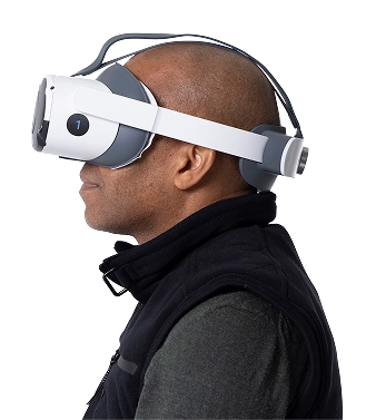
Virtual Field’s 10-2 exam is faster, easier, and more accessible than testing on traditional tabletop perimeters. As a portable 10-2 perimeter, Virtual Field allows you to perform this detailed macular exam in any exam room or mobile setting—eliminating the need for bulky equipment or darkened rooms. Our headset doesn’t require much space or a darkened room. Virtual Field can be used almost anywhere in your practice — wherever the patients feel comfortable. This way, you can improve the exam experience, expand your practice’s service offerings, and provide more comprehensive eye care to more of your patients.
Academic references and clinical validation
This study found that 10-2 visual field tests often detect central visual field defects that may be missed by 24-2 tests in glaucoma patients, glaucoma suspects, and ocular hypertensives. Abnormal results on the 10-2 test were identified in 61.5% of early glaucoma cases, 39.5% of glaucoma suspects, and 35.4% of ocular hypertensives classified as normal by 24-2 testing.
The Virtual Field macular 10-2 test provides high-resolution results that correlate well with OCT imaging, offering greater diagnostic clarity especially in glaucoma suspects and early-stage disease.
A recent study compared the 24-2, 24-2C, and 10-2 visual field tests to determine when to incorporate the 10-2. The results suggest that when 24-2 testing shows poorer mean deviation or more central defects, the 10-2 test can offer more detailed insights that help monitor disease progression.
The 24-2C and 10-2 visual field tests perform similarly in detecting overall central vision loss, but the 10-2 stands out by identifying more detailed defect clusters and aligning better with optical coherence tomography (OCT) imaging. While the 24-2C is quicker and more effective for spotting central defects, the 10-2 offers a more thorough look at central vision changes.
Did you know? Virtual Field includes automated audio translations in over 40 languages for inclusive patient engagement?
.avif)
30 days free.
No strings attached.
We are confident you’ll love Virtual Field just like the 2,000 doctors who have already made the switch.
The 10-2 Visual Field Exam at a Glance
Pros and cons of the 10-2 Exam
The pros and cons that follow can help guide you toward the ideal scenarios to incorporate this test into your patients' diagnostic assessments.
Pros
This exam offers a high-resolution assessment of the central vision field.
Fine testing detail means you can catch vision changes and intervene sooner.
The 10-2 is indispensable for monitoring macular and optic nerve conditions, especially in the earliest stages.
This exam can be combined with others, like the 24-2 and 30-2, for a more complete map of the patient’s visual field.
Cons
The 10-2 is limited to the central 10 degrees of vision, so additional tests will be required to evaluate the full visual field.
With older equipment, the 10-2 exam can feel long and stressful to patients.
Like other visual field tests, collecting accurate results depends on patient focus and cooperation.
The 10-2 visual field exam is extremely sensitive, so even small visual defects may register as abnormal results. This could lead to false positives.
List of Ocular Diseases Monitored and Diagnoses Identified by the 10-2 Visual Field Exam
Glaucoma
Glaucoma is a leading cause of irreversible blindness, and patients may miss early-stage symptoms. The 10-2 visual field test can pick up on some of the most minute changes to central vision, so many ophthalmologists offer this test to patients who are glaucoma suspects.
Neurological Disorders
Stroke, brain tumors, TBI, and pituitary disorders can all affect the optic nerve or optic chiasm. The 10-2 can help identify changes in the visual field following a cerebrovascular event or other neurological condition.
Diabetic Retinopathy
Diabetic retinopathy damages blood vessels in the retina, causing bleeding, swelling, and visual field defects. Specifically, macular edema and ischemia can lead to central vision loss that can be identified with the 10-2 test.

60+
Macular Degeneration is the leading cause of vision loss in Americans aged 60 and older
2.5M
AMD affects 2.5 million Canadians
Optic Neuropathy
Inflammation of the optic nerve, non-arteritic anterior ischemic optic neuropathy, and other neuropathies can cause sudden loss of central vision, which can be identified and confirmed with a 10-2 test. Whether through medication, nutrition, or other exposure, central vision loss due to toxic optic neuropathy can be difficult to diagnose without then 10-2 visual field test. This test is also useful in Plaquenil visual field monitoring, where early central vision changes may indicate drug toxicity long before structural damage is visible.
Macular Degeneration
Blurriness, dark spots, and distortion of the central visual field point to age-related macular degeneration. The 10-2 test can identify and monitor these symptoms, especially in the early stages when structural changes in the retina may still be undetectable in other tests.
Multiple Sclerosis (MS)
MS can cause demyelination in the central nervous system and lead to optic neuritis or lesions. In fact, research shows that nearly all patients with MS will have abnormal visual field results — but not all will notice the gradual changes in vision. The increased sensitivity of the 10-2 exam can help monitor disease progression.
Other Conditions
The 10-2 visual field exam can also monitor:
- Retinal vein/artery occlusion
- Retinal detachment
- Retinitis pigmentosa
- Autoimmune and inflammatory disorders
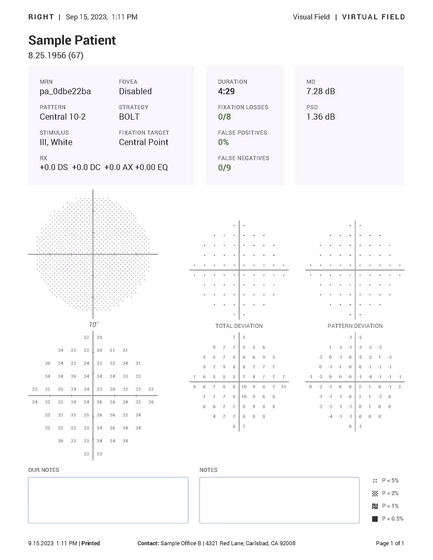
Example 10-2 Report
30 days free.
No strings attached.
We are confident you’ll love Virtual Field just like the 2,000 doctors who have already made the switch.

Billing and Coding for the 10-2 Visual Field Exam
The 10-2 visual field exam is billable to insurance under CPT code 92083, which is designated for extended visual field exams.
According to the Medicare Physician Fee Schedule (MPFS), reimbursement usually ranges from $40 to $90 per test. Your exact fees will depend on your location, setting, and other payer-specific factors.
When is the 10-2 visual field exam required?
The 10-2 visual field exam is particularly useful for glaucoma, macular degeneration, diabetic retinopathy, neurological conditions, trauma-related vision loss, and toxic retinopathy. For patients with moderate to advanced glaucoma or those showing central visual field defects on a 24-2 exam, the 10-2 can be conducted every three to six months to track progression.
This test may also be ordered for individuals taking medications like hydroxychloroquine or people with diabetes, hypertension, and neurological conditions that increase the risk of central vision loss. The 10-2 test is best used to detect subtle changes in the macular region that patients may not notice on their own. It is also a valuable tool for Plaquenil visual field monitoring, helping detect early signs of retinal toxicity in patients taking hydroxychloroquine.
Is the 10-2 visual field test required for driver’s licenses?
In most U.S. states and Canada, basic field-of-vision checks are all that’s required for driver’s licensing. The 10-2 is a more advanced, targeted screening, so it’s not mandatory for drivers.
Start Conducting 10-2 Visual Field Exams with Virtual Field
The 10-2 visual field test is an opportunity to diagnose and monitor a range of eye and neurological conditions, especially when broader tests are inconclusive. More data points in the central 10 degrees can reveal the earliest signs of glaucoma, help diagnose neurological conditions and neuropathies, and monitor macular degeneration with better precision.
You can improve the patient experience and collect pinpoint accurate data with Virtual Field. As a portable 10-2 perimeter and central visual field device, Virtual Field simplifies the 10-2 visual field test workflow — making it faster, more efficient, and far more accessible to your most vulnerable patients.
Want all 23 exam guides in one place?
Download our comprehensive guide for 160+ pages of insights.

FAQs
1. How long does a 10-2 visual field take on a head-mounted perimeter?
2. Why is the 10-2 pattern preferred for Plaquenil® toxicity monitoring?
3. Which CPT code covers a 10-2 exam?
4. Does Virtual Field record fixation losses and false-positives?
5. What mean deviation (MD) change is clinically significant on 10-2?
6. Can I export 10-2 results to my EHR?
Ready to get started?
Schedule a demo or begin your 30-day free trial of Virtual Field to try our EOM exam in your practice.
Questions? Contact sales@virtualfield.io talk to a Virtual Field expert today.
The 10-2 visual field exam pinpoints abnormalities in the central vision, which is, of course, essential for everyday tasks like reading and driving. These tasks may become more difficult for patients, but they might not attribute it to vision changes before symptoms become bothersome.
This test’s precision is key to monitoring progressive eye conditions and detecting early symptoms. Still, this supplemental test can feel time-consuming to patients; it doesn’t offer a full-scope review of the patient’s visual field, and because it’s so sensitive, there may be a higher risk of false positives.