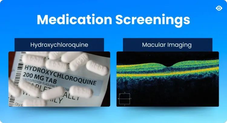
Hydroxychloroquine, commonly known by the brand name Plaquenil, remains a cornerstone therapy for autoimmune and inflammatory diseases such as rheumatoid arthritis, systemic lupus erythematosus (SLE), and various dermatologic conditions. Its efficacy and relatively low cost make it a widely prescribed medication. However, despite its benefits, hydroxychloroquine carries a critical risk of retinal toxicity, which can lead to irreversible vision loss if not detected early.
Hydroxychloroquine Eye Test: Plaquenil Eye Exam Guidelines for 2025
What Is Hydroxychloroquine Toxicity?
Hydroxychloroquine (HCQ), also known by the brand name Plaquenil, is a medication primarily used to treat autoimmune diseases such as rheumatoid arthritis, lupus, and also for malaria prevention. While effective, long-term or high-dose use of HCQ can lead to serious adverse effects, collectively known as hydroxychloroquine toxicity.
Hydroxychloroquine Toxicity Symptoms
For thise experiencing hydroxychloroquine toxicity, most early toxicity is asymptomatic, but when symptoms do develop, they may include:
- Paracentral scotomas (small blind spots near the center of vision)
- Mild contrast sensitivity loss
- Reduced night vision
- Subtle color vision changes
When the condition has progressed, or a patient developes progressive or late-stage toxicity, symptoms may include:
- Central vision loss
- Difficulty reading or recognizing faces
- Trouble seeing in dim light (nyctalopia)
- Ring scotoma (peripheral blind spot surrounding central vision)
- Visual field constriction
One of the key manifestations of hydroxychloroquine toxicity is retinal toxicity (Hydroxychloroquine Retinopathy).
What is Hydroxychloroquine Retinopathy?
Hydroxychloroquine retinopathy, often termed Plaquenil toxicity, is characterized by damage primarily to the retinal photoreceptors and the retinal pigment epithelium (RPE). This toxicity is cumulative and dose-dependent, with the daily dosage relative to real body weight being the most significant modifiable risk factor. Studies indicate that maintaining a dosage below 5 mg/kg/day significantly reduces the risk of toxicity, with less than 1% incidence within the first 5 years and under 2% risk up to 10 years of treatment [1].
Toxicity is typically irreversible and progressive, even after discontinuation of the drug, making early detection through screening vital to preserving central vision and patient quality of life [2].
Pathophysiology and Clinical Presentation of Plaquenil Retinopathy
Hydroxychloroquine accumulates in retinal cells, causing metabolic disruption and damage at the level of photoreceptors, particularly in the outer nuclear layer, followed by secondary changes in the RPE [3]. The classic clinical hallmark is a bilateral bull’s-eye maculopathy, marked by a ring-shaped area of parafoveal depigmentation sparing the foveal center, more commonly seen in patients of European, African-American, and Hispanic descent.
Interestingly, patients of Asian descent often present with early toxicity changes in a more peripheral, extramacular distribution near the vascular arcades, necessitating adjusted screening techniques to avoid missed diagnoses [4]. Importantly, toxicity may progress even after drug cessation due to delayed retinal cell death, underscoring the need for vigilant long-term follow-up.
Key Risk Factors for Retinal Toxicity in Plaquenil Users
Several key factors influence the risk of hydroxychloroquine toxicity:
- Daily dosage exceeding 5 mg/kg of real body weight
- Duration of therapy longer than 5 years
- Renal impairment reducing drug clearance
- Concurrent use of tamoxifen, which may potentiate retinal toxicity
- Pre-existing macular or retinal disease
- Older age and cumulative lifetime dose
- Genetic predispositions and retinal pigment variations in certain ethnic groups
Regular monitoring and dose adjustments are essential to mitigate these risks.
How to Conduct a Hydroxychloroquine (Plaquenil) Eye Exam
The primary goal of screening for Plaquenil toxicity is to identify early retinal changes before functional vision loss occurs, enabling timely discontinuation of the drug. The American Academy of Ophthalmology (AAO) recommends the following:
- Baseline eye exam within the first year of starting hydroxychloroquine, including assessment of ocular health and establishing reference imaging and functional tests.
- Annual screening after 5 years of continuous use, or earlier if risk factors are present.
- More frequent exams if patients have renal impairment, use tamoxifen, or have existing retinal pathology.
While baseline functional testing such as automated visual fields and spectral domain optical coherence tomography (SD-OCT) is helpful, it is most critical once therapy is ongoing and risk increases.
Diagnostic Tools for Hydroxychloroquine Toxicity
Automated Visual Field Testing
The 10-2 visual field test is the standard for detecting macular visual field defects in non-Asian patients, offering high-resolution sensitivity for parafoveal damage. For patients of Asian descent, broader patterns such as 24-2 or 30-2 fields are advised to capture peripheral lesions.
Visual field testing remains somewhat subjective and can be affected by patient reliability; thus, confirmatory objective testing is recommended before diagnosing toxicity.
Spectral Domain Optical Coherence Tomography (SD-OCT)
SD-OCT provides a non-invasive, high-resolution cross-sectional view of retinal layers. Early toxicity manifests as localized thinning or disruption of the photoreceptor outer segments and the ellipsoid zone, often appearing as focal defects in the parafoveal area for non-Asian patients or near the arcades in Asian patients.
These findings are highly specific and often precede visual field changes, making SD-OCT indispensable in screening.
Fundus Autofluorescence (FAF) Imaging
FAF highlights metabolic stress in the RPE by detecting lipofuscin accumulation. Early Plaquenil toxicity may appear as areas of increased autofluorescence, sometimes even before structural changes are visible on SD-OCT. Late-stage toxicity causes hypoautofluorescence, indicating RPE atrophy.
Multifocal Electroretinography (mfERG)
MfERG objectively measures localized retinal electrical activity and is valuable in corroborating functional deficits noted on visual fields. It can detect subtle regional dysfunction before structural damage is apparent.
For more on vital eye care technology, review this roadmap to enhanced patient outcomes and practice success.
Interpreting Screening Results and Clinical Decision-Making
Diagnosis of Plaquenil toxicity is typically made based on a combination of subjective functional tests (visual fields) and objective structural tests (SD-OCT, FAF, mfERG). Ambiguous or borderline findings warrant repeat testing to confirm consistency and avoid false positives.
Once toxicity is confirmed, the ophthalmologist should promptly communicate with the prescribing physician to consider discontinuation of hydroxychloroquine or dosage reduction. Continued monitoring is essential, as retinal damage may progress despite stopping the drug.
Patient Education and Long-Term Management
Patients on hydroxychloroquine should be educated about the importance of adherence to screening schedules and reporting new visual symptoms such as difficulty reading, blurring, or changes in color vision. Encouraging lifestyle factors to support retinal health, including smoking cessation and UV protection, may also be beneficial.
Given the expanding use of hydroxychloroquine in multiple specialties, interdisciplinary collaboration between rheumatologists, dermatologists, and eye care providers is critical to optimize therapy and preserve vision.
How Virtual Field Improves Screening for Hydroxychloroquine Toxicity
Virtual Field provides significant improvements in screening for hydroxychloroquine toxicity, especially when compared to traditional bowl perimeters and manual testing workflows. Because early detection is critical in preventing irreversible vision loss from Plaquenil retinopathy, doctors and specialists need flexibility, progression monitoring, and comfortable exam experiences that ensure routine patient testing and compliance.
Virtual Field supports both protocols 10-2 and 24-2, which are the exams recommended by the AAO's screening guidelines for the early detection of hydroxychloroquine toxicity, and can be performed without bulky equipment. In addition, Virtual Field testing:
- Can be conducted in any exam room
- Enables testing for patients with limited mobility, which is especially important for patients with autoimmune conditions or older populations
- Encourages more frequent and consistent screening by reducing practice workflow frictions and patient testing anxiety
Conclusion
Hydroxychloroquine remains an invaluable medication for many patients but carries a small, significant risk of irreversible retinal toxicity. Early and regular screening using a multimodal approach involving visual fields, SD-OCT, FAF, and mfERG is essential to detect preclinical changes. By adhering to established guidelines and understanding risk factors, eye care professionals can safeguard patients' vision while allowing them to benefit from this important therapy.
References
- Melles RB, Marmor MF. The risk of toxic retinopathy in patients on long-term hydroxychloroquine therapy. JAMA Ophthalmol 2014;132:1453–60.
- Marmor MF, Hu J. Effect of disease stage on progression of hydroxychloroquine retinopathy. JAMA Ophthalmol 2014;132:1105–12.
- Marmor MF. Comparison of screening procedures in hydroxychloroquine toxicity. Arch Ophthalmol 2012;130:461–9.
- Melles RB, Marmor MF. Pericentral retinopathy and racial differences in hydroxychloroquine toxicity. Ophthalmology 2015;122:110–6.
- Marmor MF et al. Recommendations on screening for chloroquine and hydroxychloroquine retinopathy (2016 Revision). Ophthalmology 2016;123:1386-94.
About Virtual Field
Virtual Field delivers an exceptional eye exam experience. Eye care professionals including ophthalmologists and optometrists examine patients faster, more efficiently, and more comfortably than ever before. Exams include Visual Field, 24-2, Kinetic Visual Field (Goldmann Perimetry), Ptosis, Esterman, Color Vision, Pupillometry, Extraocular Motility (EOM), and more.


