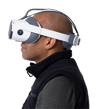
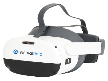
Most eye exams measure vision using high-contrast stimuli, but that doesn’t tell the whole story. The visual contrast sensitivity test captures the nuance of how vision performs in low-light, low-contrast, or high-glare conditions. This often underutilized test is relevant for patient quality of life and can help diagnose progressive conditions at their earliest stages.
Many patients with poor contrast sensitivity still have normal acuity on standard charts. A low contrast vision assessment uncovers deficits in real-life conditions like night driving or reading in dim light. A modern contrast sensitivity test device helps eye care providers detect these problems early — when intervention is most effective.
Visual acuity and color vision testing may be leveraged more frequently, but the contrast sensitivity eye exam deserves more focus.
How a Contrast Sensitivity Test Device Measures Low Contrast Vision
Contrast sensitivity testing is increasingly recognized as a valuable tool for evaluating visual function, but it is still not a core component of most routine eye exams. Traditionally, contrast sensitivity is measured using printed sine-wave gratings or contrast charts, such as the Pelli-Robson, Bailey-Lovie contrast chart, and the Mars Letter test. These tests determine the minimum level of contrast a patient can detect. However, these paper-based methods can fade, be inconsistently lit, or suffer from observer bias.
Academic references and clinical validation
A recent survey in the British Journal of Ophthalmology found that, for patients with age-related macular degeneration, contrast sensitivity has a stronger correlation with vision-related quality of life than visual acuity.
Changes in contrast sensitivity could indicate damage to the retina. Conducting this exam could help lead to earlier diagnoses of diabetic retinopathy.
Tablet-based and virtual reality contrast sensitivity testing methodologies have been shown to be as effective and reliable as traditional methods.
Research increasingly supports transitioning from paper-based tools to a contrast sensitivity test device that reduces bias, offers standardized luminance, and enables consistent low contrast vision assessment across environments.
Did You Know?
The average Virtual Field customer gets an extra 62 working hours back thanks to the workflow efficiencies from Virtual Field.
.avif)

30 days free.
No strings attached.
We are confident you’ll love Virtual Field just like the 2,000 doctors who have already made the switch.
Low Contrast Vision Assessment: What to Expect from the Test
It’s essential to distinguish visual contrast sensitivity testing from visual acuity and visual field exams. In this exam, patients are challenged to detect subtle patterns at varying contrast levels. They may notice a reduction in “sharpness” in their daily life, but often, patients don’t realize how much their functional vision has declined until they struggle with this test.
Eye care professionals sometimes skip this test because it can produce imprecise results using traditional methods, but it is a valuable diagnostic tool that should not be overlooked.
Using a contrast sensitivity test device with pre-calibrated settings eliminates the inconsistencies often found in older contrast charts and offers a standardized baseline for diagnosing retinal dysfunction.
Pros and Cons of Using a Contrast Sensitivity Test Device
The pros and cons that follow can help guide you toward the ideal scenarios to incorporate this test into your patients' diagnostic assessments.
Pros
A modern contrast sensitivity test device reveals visual challenges undetected by acuity-only exams.
Its results have real-world applications that can improve patient quality of life.
Results can indicate the early stages of common conditions, such as AMD or diabetic retinopathy.
Devices like a portable CSV-1000 alternative simplify the process and can integrate into routine vision assessments.
Cons
Contrast sensitivity is not a standard component of routine screenings. Therefore, it may add time to your workflows.
Traditional, manual methods can provide subjective test results.
Contrast sensitivity testing cannot be billed separately.
Patients can take a contrast sensitivity test online, which can yield mixed results.
Many providers still rely on outdated tools; those looking to buy a contrast sensitivity chart should seek calibrated or device-based options that meet modern testing standards.
Eye Conditions Identified by Low Contrast Sensitivity Testing
A low contrast vision assessment helps uncover functional vision loss that may occur early in a variety of conditions, including cataracts, diabetic retinopathy, and neuro-ophthalmic disease.
Cataracts
Even early-stage cataracts can scatter light and degrade contrast sensitivity. This test can be more sensitive than acuity charts in early detection. These subtle changes are often missed without a contrast sensitivity test device, especially in early stages.
Diabetic Retinopathy
Patients with diabetes may report foggy or “washed-out” vision even when their acuity remains good. Contrast testing helps explain this disconnect and can help identify early stages of diabetic retinopathy.

1 in 6
Roughly 1 in 6 U.S. adults over 40 already has at least one cataract
> 3 million
Cataract surgeons perform >3 million cataract procedures
per year
Glaucoma
As retinal ganglion cells die off, especially in the early stages of glaucoma, contrast sensitivity can decrease before noticeable vision loss is detected on a visual field test.
Age-Related Macular Degeneration (AMD)
Difficulty distinguishing fine details can be the first sign of AMD. It can also signal changes in the retinal pigment epithelium. In both cases, contrast sensitivity testing can help inform management techniques.
Multiple Sclerosis
MS can impact the optic nerve, leading to reduced contrast even when central acuity is unaffected. It’s also helpful in tracking recovery post-optic neuritis.
30 days free.
No strings attached.
We are confident you’ll love Virtual Field just like the 2,000 doctors who have already made the switch.

Billing Codes and Charting for Contrast Sensitivity Vision Assessments
Contrast sensitivity is not an independently billable test, but it can be documented as part of a sensorimotor or functional assessment. Depending on the context, you can bill it under CPT code 92060 or 92283. Review the Medicaid Fee Schedule in your state for the latest definitions and details.
Documenting contrast sensitivity results from a portable CSV-1000 alternative or digital testing system can support broader functional vision assessments.
When is the contrast sensitivity test required?
Patients with complaints of poor night vision, glare issues, or visual discomfort in low-contrast settings should be tested for contrast sensitivity. They may have vague complaints of their vision feeling foggy or faded. This sensation becomes more common with age, so patients at high risk of cataracts, glaucoma, AMD, or diabetic retinopathy should be tested. Contrast sensitivity can temporarily drop after LASIK or PRK, so regular testing helps measure patient recovery.
People with certain professions, such as pilots or machine operators, may need vision contrast sensitivity testing to meet occupational requirements. For other patients, it can be a matter of everyday safety.
Is contrast sensitivity testing required for driver’s licenses?
Surprisingly, no, the U.S. Department of Transportation doesn’t mandate contrast sensitivity testing for standard driver’s licensing. Although low contrast sensitivity can be highly hazardous to drivers, most states rely on visual acuity and visual field testing for licensing. Results from the Esterman exam or FullField 120 are usually sufficient.
Complete Your Comprehensive Exams with Virtual Field
Early contrast sensitivity issues can signal vision decline well before acuity drops. Adding a contrast sensitivity test device to your workflow allows for efficient, accessible low contrast vision assessments for all at-risk patients. For patients at risk of AMD or diabetic retinopathy, you can add this test to your screening routines without disrupting your workflow.
Virtual Field’s visual field tests can provide more insight and help monitor disease progression. You can add Virtual Field to your testing routines for comprehensive, precise, and patient-friendly eye exam experiences on a larger scale.
Want all 23 exam guides in one place?
Download our comprehensive guide for 160+ pages of insights.
FAQs
1. Why measure contrast sensitivity when Snellen acuity is 20/20?
2. Is there a dedicated CPT code for contrast sensitivity?
3. What constitutes a normal Pelli-Robson score?
4. Can results predict visual quality after IOL implantation?
5. Why doesn't Virtual Field offer a Contrast Sensitivity test?
Ready to get started?
Schedule a demo or begin your 30-day free trial of Virtual Field to try our EOM exam in your practice.

Questions? Contact sales@virtualfield.io talk to a Virtual Field expert today.


