
What is a Pupillometry Exam, and What Does it Measure?
A pupillometry exam involves the use of a pupillometer — a specialized instrument designed to precisely measure the size, shape, and reactivity of the pupils. This non-invasive exam evaluates how pupils respond to light stimuli, providing critical insights into the health of the optic nerve, retina, and neurological pathways.
Key components assessed in a comprehensive pupillometry exam include:
- Integrity of the pupillary light reflex pathway
- Detection of light-near dissociation
- Identification of relative afferent pupillary defects (RAPD)
Pupil Structure and the Role of Pupillometry
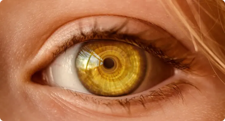
The pupil, located centrally within the iris, regulates the amount of light entering the eye. Its dynamic size changes — from constricted to dilated — allow optimal vision under varying light conditions. These size adjustments are governed by intricate neural pathways linking the retina, optic nerve, and brainstem.
A pupillometer precisely quantifies these responses, providing objective data on pupil size and reaction times. This data is invaluable in detecting subtle dysfunctions that might be missed by traditional manual exams.
Traditional Pupillometry vs. Automated Pupillometer Exams
Historically, pupillary exams involved the swinging flashlight test, where clinicians observed the pupil’s reaction to a light source subjectively. This method was prone to variability and often required expert interpretation.
With the advent of automated pupillometers, pupillometry exams have become standardized and reproducible. These devices employ infrared videography or computerized sensors to capture detailed pupil responses quickly and accurately. Infrared-based pupillometers are particularly effective for patients with dark irises, as infrared light reflects off melanin to enhance pupil visibility.
Advantages of Automated Pupillometry Using a Pupillometer
Automated pupillometry offers multiple advantages over traditional methods:
- Objectivity: Removes human error and observer bias.
- Speed: Provides immediate and precise measurements.
- Ease of Use: Can be performed by trained technicians before dilation or provider examination.
- Consistency: Enables reliable longitudinal tracking of pupil function.
These benefits make automated pupillometer exams a staple in many eye clinics, neurology departments, and intensive care units.
Virtual Reality Technology: The Future of Automated Pupillometry Testing
Emerging virtual reality (VR) technology is further transforming automated pupillometry exams by creating immersive and highly controlled testing environments. VR-based pupillometry allows eye care professionals to precisely monitor pupil responses in real time while patients interact with dynamic visual stimuli within a headset.
Additionally, VR-based pupillometry can seamlessly integrate with other diagnostic tests such as visual field assessments, enabling a comprehensive evaluation of visual and neurological function in one session. The portability and flexibility of VR headsets also facilitate testing in diverse clinical environments, from primary eye care offices to research labs and even remote telehealth setups.
By combining automated pupillometry with immersive VR, clinicians gain richer, multidimensional data on pupil behavior, improving early detection of neurological disorders, glaucoma progression, and postoperative visual outcomes. As this technology evolves, VR-enabled pupillometry promises to be a powerful tool in precision eye care and neuro-ophthalmology.
Clinical Applications of Pupillometry Exams
Pupillometry is a powerful diagnostic tool used in a variety of clinical scenarios:
- Neurological Assessment: Detecting relative afferent pupillary defects (RAPD) to diagnose optic nerve damage from ischemia, compression, or demyelinating diseases.
- Glaucoma Screening: Integrated with visual field testing to detect early optic nerve dysfunction.
- Preoperative LASIK Evaluation: Automated pupillometers assess pupil size to predict night vision disturbances post-surgery, as recommended by the American Society of Cataract and Refractive Surgery (ASCRS).
- Critical Care Monitoring: In the ICU, pupillometry helps detect neurological deterioration in traumatic brain injury patients by tracking changes in pupil reactivity and size that precede complications like brain herniation.
Understanding Relative Afferent Pupillary Defect (RAPD) through Pupillometry
One of the hallmark findings detected by automated pupillometry is the presence of RAPD, an asymmetry in pupil response caused by optic nerve or severe retinal disease. A pupillometer provides quantitative measurements to identify RAPD with high sensitivity, often earlier than clinical symptoms appear.
Read our white paper by Dr. Lisa Arbisser to learn about Pupillometry and its uses in diagnosing neurological conditions.

Billing for Automated Pupillometry Exams
Reflecting its growing clinical importance, a dedicated CPT code (95919) for quantitative automated pupillometry was introduced in January 2023. Eye care providers can now bill for pupillometer exams, provided a licensed practitioner interprets and documents the findings for unilateral or bilateral measurements.
This billing advancement emphasizes how automated pupillometry is no longer a niche test but a mainstream diagnostic tool offering crucial insights into eye and neurological health.
Conclusion
Automated pupillometry and the use of modern pupillometers represent a major leap forward in eye care diagnostics. By delivering fast, accurate, and objective pupil measurements, this technology empowers clinicians to detect optic nerve and neurological conditions earlier and with greater confidence. Whether in outpatient eye clinics, surgical screening, or critical care environments, pupillometry is now an indispensable part of comprehensive patient evaluation.
For more in-depth information, check out our white paper by Dr. Lisa Arbisser on pupillometry and its role in neurological diagnosis.
About Virtual Field
Virtual Field delivers an exceptional eye exam experience. Eye care professionals including ophthalmologists and optometrists examine patients faster, more efficiently, and more comfortably than ever before. Exams include Visual Field, 24-2, Kinetic Visual Field (Goldmann Perimetry), Ptosis, Esterman, Color Vision, Pupillometry, Extraocular Motility (EOM), and more.